44 human brain diagram with labels
Brain: Ultimate Guide to the Brain for AP® Psychology - Albert The forebrain consists of the thalamus, hypothalamus, amygdala, and the hippocampus. The hypothalamus, amygdala, and hippocampus make up what we call the Limbic System of your brain. Thalamus The thalamus is located between the cerebral cortex and the midbrain. It is made up of nuclei that receive different sensory and motor inputs. Free Printable Brain Hemisphere Hat - Homeschool Giveaways The brain is not fully developed until you are 25 years old. A human brain is made up of 60% fat. Our brain's storage capacity is unlimited. More storage than a super computer. The human brain weighs 3 pounds. A brain can generate enough power to power up a lightbulb.
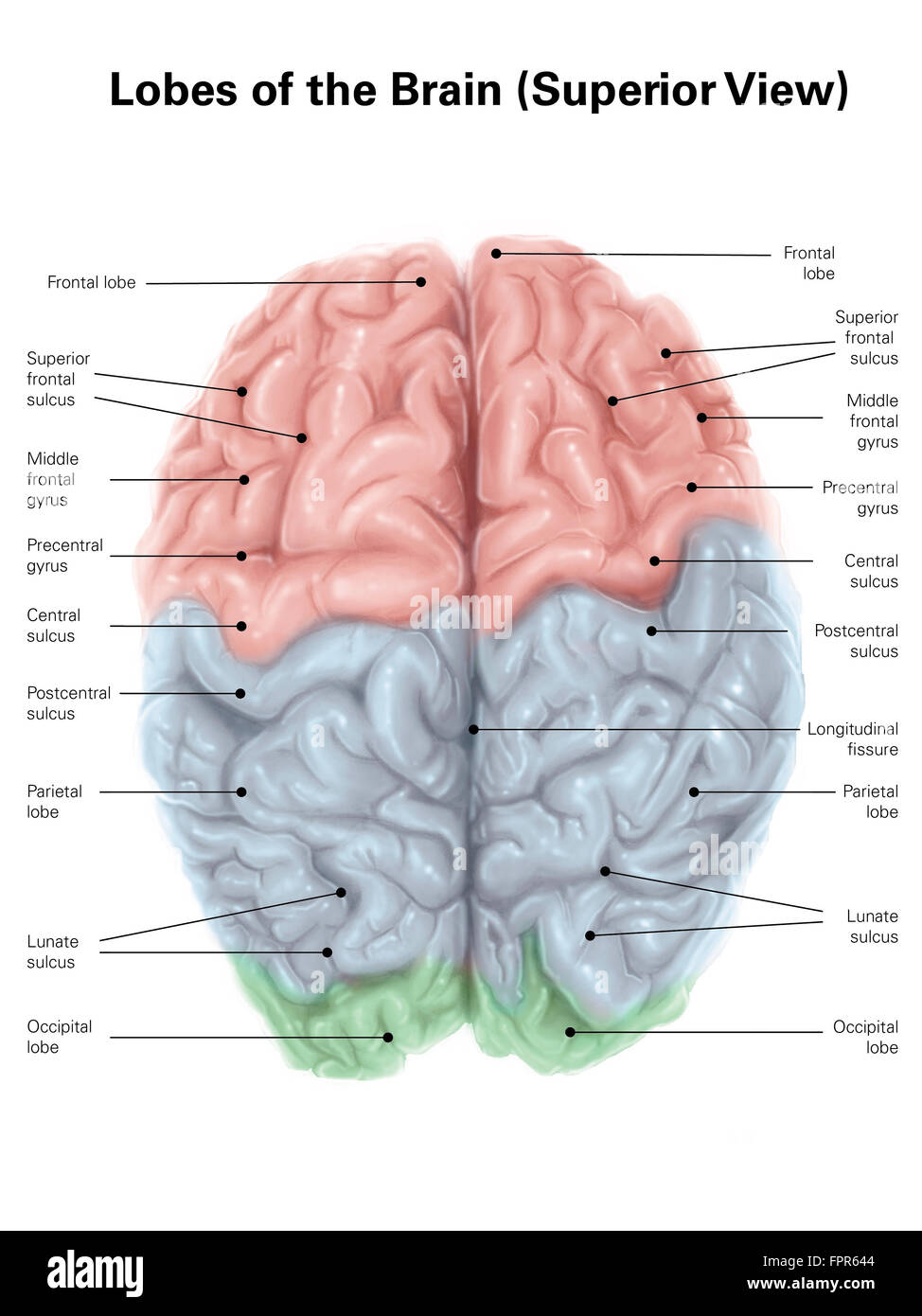
Positions and Functions of the Four Brain Lobes - MD-Health.com Composed of 50 to 100 billion neurons, the human brain remains one of the world's greatest unsolved mysteries. Here we will take a closer look at the four lobes of the brain to discover more about the location and function of each lobe. Brain Lobes and their Functions. The brain is divided into four sections, known as lobes (as shown in the image).
Human brain diagram with labels
FREE Human Body Systems Labeling with Answer Sheets The free respiratory system labeling sheet includes a blank diagram to fill in the trachea, bronchi, lungs, and larynx. The free nervous system labeling sheet includes blanks to label parts of the brain, spinal cord, ganglion, and nerves. The free muscular system labeling sheet includes a blank diagram to label some of the main muscles in the body. Left Brain vs. Right Brain: Characteristics Chart [INFOGRAPHIC] Brain dominance theory is absorbing and enjoyable, plus it allows people to think about stereotypes and labels. In reality, though, psychology is complicated, and the truth is that there are very few people who have the traits of only one of these descriptions. More often, we have an intricate combination of both. › photos › diagram-of-bodyDiagram Of Body Organs Female Pics Pictures, Images ... - iStock Human internal organs Internal organs in woman and man body. Brain, stomach, heart, kidney, medical icon in female and male silhouette. Digestive, respiratory, cardiovascular systems. Anatomy poster vector illustration. diagram of body organs female pics stock illustrations
Human brain diagram with labels. The human brain: Parts, function, diagram, and more Click on the BodyMap above to interact with a 3D model of the brain. Cerebrum The cerebrum is the front part of the brain and includes the cerebral cortex. This part of the brain is responsible for... brain | Definition, Parts, Functions, & Facts | Britannica brain, the mass of nerve tissue in the anterior end of an organism. The brain integrates sensory information and directs motor responses; in higher vertebrates it is also the centre of learning. The human brain weighs approximately 1.4 kg (3 pounds) and is made up of billions of cells called neurons. Junctions between neurons, known as synapses, enable electrical and chemical messages to be ... 14 Informative Facts, Diagram & Parts Of Human Brain For Kids Brainstem: Brainstem is the third region of the brain that anchors the spinal cord. Located in front of the cerebellum, the brainstem consists of the pons, medulla, and the midbrain. The brainstem is involved in essential life functions, such as breathing, regulation of blood pressure, and heart rate. Parts of the Brain Activity for Kids, Brain Diagram, and Worksheets for ... Human Body Printables makes a printable book that teaches students all about their heart, brain, muscles, cells, skin, bones, lungs, stomach, intestines, and bladder 4 EPIC Skeletal System Project ideas for kids - using things like pasta, life saver gummies, lego, and more you can learn about the human body in a fun memorable way
Brain Diagram Pons Images, Stock Photos & Vectors | Shutterstock brain diagram pons images 338 brain diagram pons stock photos, vectors, and illustrations are available royalty-free. See brain diagram pons stock video clips of 4 hypothalamus vector brain diagram with labels labeled brain anatomy pons human body anatomy with labels ventricles in the brain the hypothalamus midbrain cerebellum thalamus Next of 4 en.wikipedia.org › wiki › File:Human_skeleton_frontFile:Human skeleton front en.svg - Wikipedia Polski: Diagram przedstawiający ludzki układ kostny (kobieta). Português : Diagrama de um esqueleto humano feminino, vista anterior. As linhas em vermelho apontam para ossos individuais e encontram-se no singular. › en › e-AnatomyAnatomical diagrams of the brain - e-Anatomy - IMAIOS Atlas of the human brain based on colored anatomical drawings and diagrams ... These original illustrations and diagrams of the brain were created from 3D medical imaging reconstructions and then redrawn and colored using Adobe Illustrator. ... The user can select to display multiple categories of labels on the illustrations: Cerebral lobes ... Anatomy of the Brain - Simply Psychology Each cerebral hemisphere can be subdivided into four lobes, each associated with different functions. The four lobes of the brain are the frontal, parietal, temporal, and occipital lobes (Figure 3). Figure 3. The cerebrum is divided into four lobes: frontal, parietal, occipital and temporal. Frontal lobes
Brain: Atlas of human anatomy with MRI - e-Anatomy - IMAIOS Anatomy of the brain (MRI) - cross-sectional atlas of human anatomy. The module on the anatomy of the brain based on MRI with axial slices was redesigned, having received multiple requests from users for coronal and sagittal slices. The elaboration of this new module, its labeling of more than 524 structures on 379 MRI images in three different ... Parts of the brain: Learn with diagrams and quizzes | Kenhub Labeled brain diagram First up, have a look at the labeled brain structures on the image below. Try to memorize the name and location of each structure, then proceed to test yourself with the blank brain diagram provided below. Labeled diagram showing the main parts of the brain Blank brain diagram (free download!) en.wikipedia.org › wiki › Human_eyeHuman eye - Wikipedia The human eye is a sensory organ, part of the sensory nervous system, that reacts to visible light and allows us to use visual information for various purposes including seeing things, keeping our balance, and maintaining circadian rhythm. The eye can be considered as a living optical device. Parts of the Human Brain | Anatomy & Function - Study.com The four lobes of the cerebrum are shown in the diagram. The cerebrum, a part of the brain, is composed of four lobes The cerebrum controls a wide range of functions, including memory, speech and...
Labeling Theory - Simply Psychology Labeling theory is an approach in the sociology of deviance that focuses on the ways in which the agents of social control attach stigmatizing stereotypes to particular groups, and the ways in which the stigmatized change their behavior once labeled. Labeling theory is associated with the work of Becker and is a reaction to sociological ...
PARTS OF THE BRAIN - The Human Memory The human brain is hugely interconnected but three major components can be identified: the cerebrum, the cerebellum and the brain stem. The brainstem which includes the medulla, the pons and the midbr ain, controls breathing, digestion, heart rate and other autonomic processes, as well as connecting the brain with the spinal cord and the rest ...
Diagram of Human Heart and Blood Circulation in It Four Chambers of the Heart and Blood Circulation. The shape of the human heart is like an upside-down pear, weighing between 7-15 ounces, and is little larger than the size of the fist. It is located between the lungs, in the middle of the chest, behind and slightly to the left of the breast bone. The heart, one of the most significant organs ...
Brain: Function and Anatomy, Conditions, and Health Tips releasing hormones Brain diagram Use this interactive 3-D diagram to explore the brain. Anatomy and function Cerebrum The cerebrum is the largest part of the brain. It's divided into two halves,...

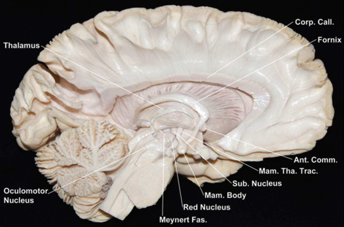
Medial Surface of the Left Hemisphere and Brainstem | Neuroanatomy | The Neurosurgical Atlas, by ...
What Is a Neuron? Diagrams, Types, Function, and More Equal numbers of neuronal and nonneuronal cells make the human brain an isometrically scaled-up primate brain. pubmed.ncbi.nlm.nih.gov/19226510/ Brain basics: The life and death of a neuron.
byjus.com › biology › liver-diagramLiver Diagram with Detailed Illustrations and Clear Labels Anatomically, the liver is a meaty organ that consists of two large sections called the right and the left lobe. The rib cage partly protects the liver and cannot be felt if you were to touch it. However, it can be felt ascending and descending if you were to take a deep breath. The liver weighs an average of 1.3 kilograms in an adult human.

Brain Anatomy Poster - Laminated - Anatomical Chart of The Human Brain- Buy Online in Guernsey ...
Body Cavities and Membranes: Labeled Diagram, Definitions Body Cavities Labeled Diagram: The dorsal cavity is located in the back of the body (red/stars) and houses the central nervous system including the brain and spinal cord. Ventral Cavity The ventral cavity is the cavity located in the front of the body, which makes sense because ventral means front or anterior.
What are the 12 cranial nerves? Functions and diagram Scientists use Roman numerals from I to XII to label the cranial nerves in the brain. The 12 cranial nerves include the: olfactory nerve optic nerve oculomotor nerve trochlear nerve trigeminal...
Human Brain Lesson for Kids: Function & Diagram - Study.com It even helps you understand what's going on around you by receiving messages from your senses: touch, taste, smell, sight, and hearing. The Cerebellum Another part of your brain is called the...
Human brain - Wikipedia The human brain is the central organ of the human nervous system, and with the spinal cord makes up the central nervous system.The brain consists of the cerebrum, the brainstem and the cerebellum.It controls most of the activities of the body, processing, integrating, and coordinating the information it receives from the sense organs, and making decisions as to the instructions sent to the ...
Human Body Anatomy Basics No Lines Clip Art at Clker.com - vector clip art online, royalty free ...
› male-human-anatomy-diagramMale Human Anatomy Diagram Pictures, Images and Stock Photos Human anatomy, back injury or disease, medical concepts. Three main curvatures of the spine disorders or deformities on male body: lordosis, kyphosis and scoliosis 3D rendering illustration. Human anatomy, back injury or disease, medical concepts. male human anatomy diagram stock pictures, royalty-free photos & images
Left and Right Hemisphere of the Brain - The Human Memory The brain has two sides and separated into unique lobes. Each lobe has a specific set of functions. Although the brain is a complex organ - a hardworking one with a hundred billion neurons, it surprisingly weighs only three pounds. It makes up around 2% of the human weight and only takes up about 20% of the body's total energy.

Draw a labeled diagram of human eye Write the functions of Cornea, Iris, Pupil, eye lens and ...
Cerebrum: Anatomy, Function, and Treatment - Verywell Health Corpus callosum: A band of white matter that joins the halves of the cerebrum at the deep center of the brain and coordinates nerve signals between each half. Cerebral arteries: Blood vessels that supply the cerebrum with oxygen-rich blood from the heart. There are three cerebral arteries: anterior (front), middle, and posterior (back).; Circle of Willis: A loop of cerebral arteries and other ...
Lobes of the brain: Structure and function - Kenhub The lobes of the cerebrum are actually divisions of the cerebral cortex based on the locations of the major gyri and sulci. The cerebral cortex is divided into six lobes: the frontal, temporal, parietal, occipital , insular and limbic lobes. Each lobe of the cerebrum exhibits characteristic surface features that each have their own functions.
Mapping the Brain to Understand the Mind - Scientific American This closeup of a single human neuron highlights just how interconnected brain cells are. False color reveals the locations and abundance of synapses where the cell receives signals from other...


Post a Comment for "44 human brain diagram with labels"