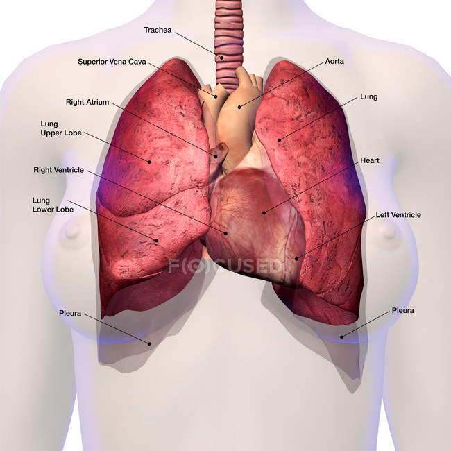
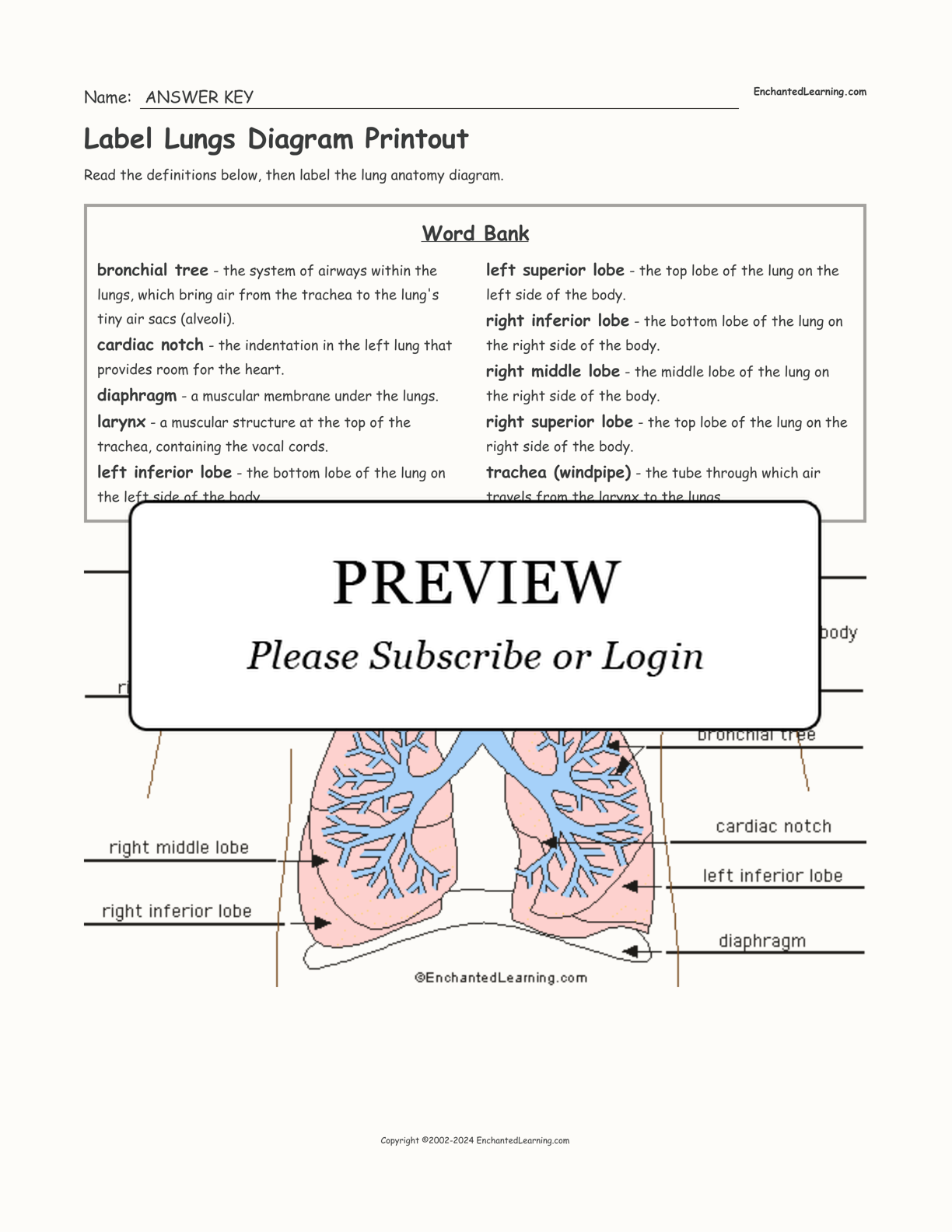
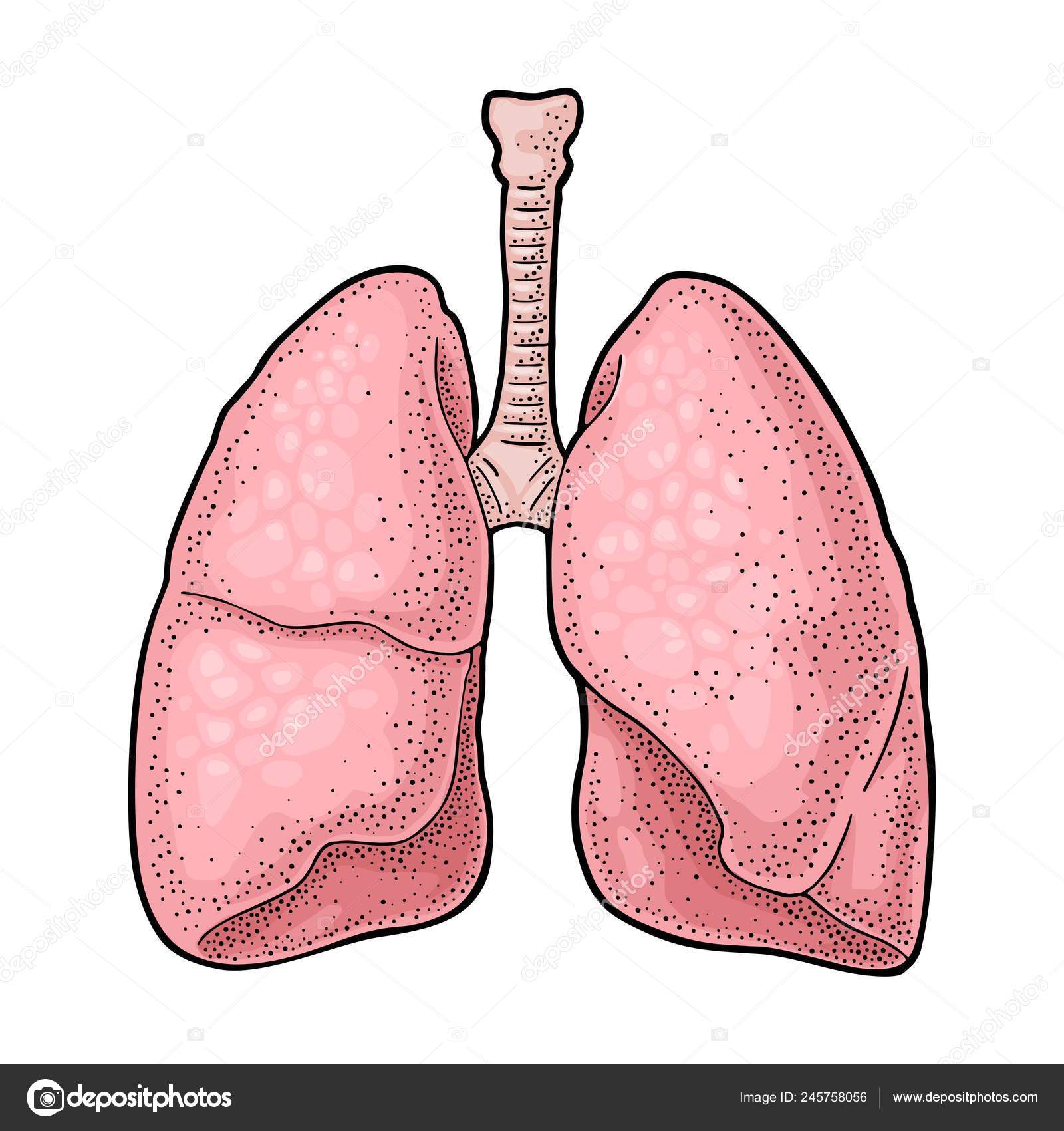
39 lungs pictures with labels
Labeled imaging anatomy cases | Radiology Reference Article ... Hacking, C. Labeled imaging anatomy cases. Reference article, Radiopaedia.org. (accessed on 22 Sep 2022) Histology, Lung - StatPearls - NCBI Bookshelf The lungs are a pair of primary organs of respiration, present in the thoracic cavity beside the mediastinum. They are covered by a thin double-layered serous membrane called the pleura. The respiratory system consists of two components, the conducting portion, and the respiratory portion. The conducting portion brings the air from outside to ...
Lung Transplant Rejection - StatPearls - NCBI Bookshelf The number of lung transplants annually in the US and worldwide has increased in recent years. This is due to the systemization of nationwide database and allocation, improved surgical techniques, and the advent of a new generation of immunosuppressants. However, lung transplantation recipients continue to have a high rate of short term and long term failure rates compared to other solid ...
Lungs pictures with labels
Lung Volumes - Definitions - Measuring - TeachMePhysiology It is useful to divide the total space within the lungs into volumes and capacities. These can be measured to aid in the definitive diagnosis, quantification and monitoring of disease. They allow an assessment of the mechanical condition of the lungs, its musculature, airway resistance and the effectiveness of gas exchange at the alveolar membrane while being, for the most part, cheap, non ... Smoker's Lungs Pictures | New Health Advisor 5 Smokers Lungs Pictures That Will Shock You If you have a weak stomach, you may not want to look at the following pictures. If you are a smoker, you will want to stop. Image 1: Lung of People Smoking for 15 Years The no.1 image is that of someone who has been smoking for 15 years. As you can see the lung has been covered in black spots. Free Heart Worksheets for Human Anatomy Lessons - Homeschool Giveaways Print out sheet of the human heart with labels - This fun heart worksheet shows kids the different parts of the heart. They'll learn about the left ventricle, the left atrium, the tricuspid valve, and more. Human Heart Clipart - There is a coloring page, heart labeling worksheet and heart anatomy chart.
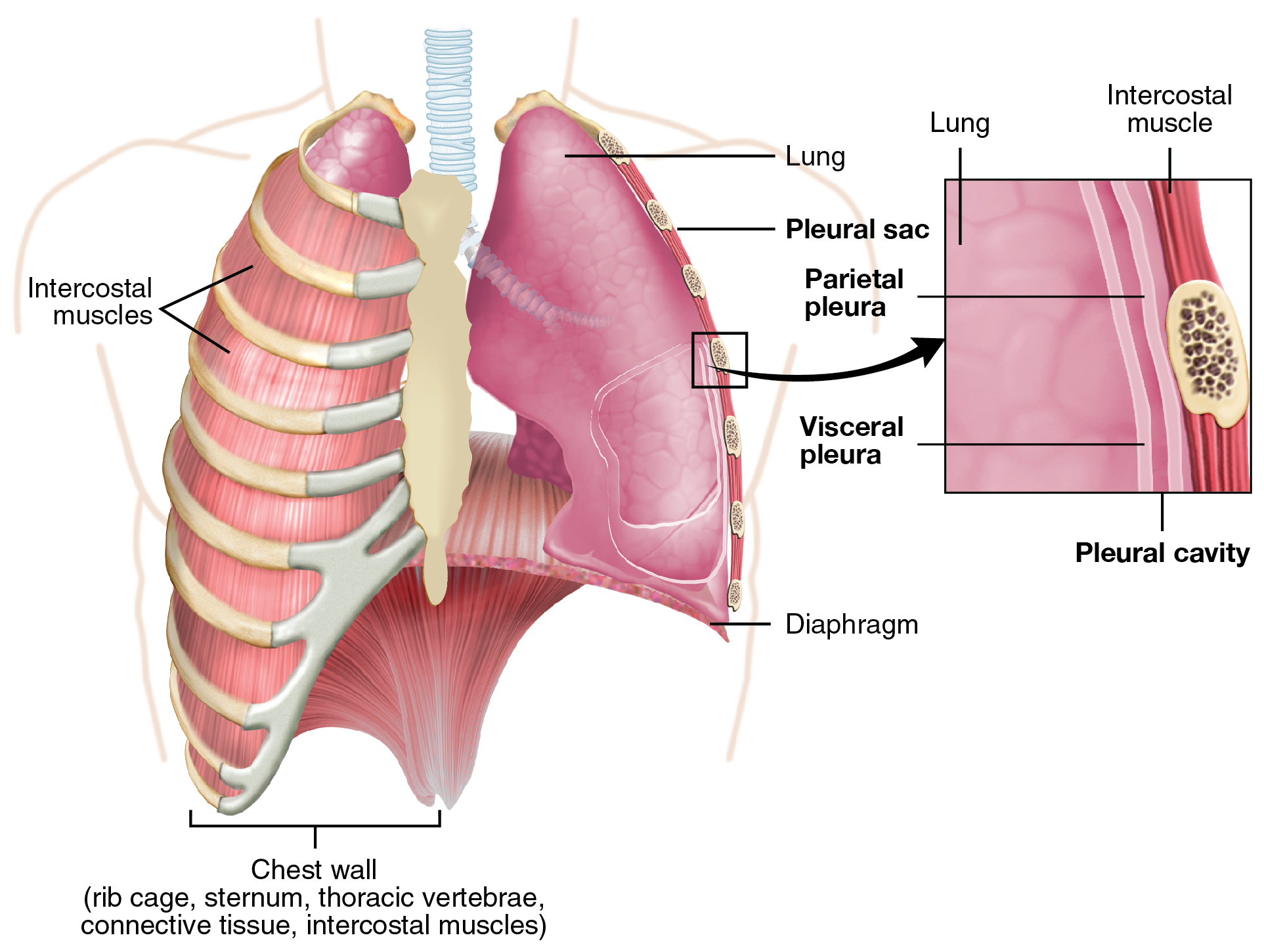
Lungs pictures with labels. Heart: illustrated anatomy - e-Anatomy - IMAIOS This interactive atlas of human heart anatomy is based on medical illustrations and cadaver photography. The user can show or hide the anatomical labels which provide a useful tool to create illustrations perfectly adapted for teaching. Anatomy of the heart: anatomical illustrations and structures, 3D model and photographs of dissection. Pleura: Anatomy, Function, and Conditions - Verywell Health The pleura is a vital part of the respiratory tract whose role it is to cushion the lungs and reduce any friction which may develop between the lungs, rib cage, and chest cavity. The pleura consists of a two-layered membrane that covers each lung. The layers are separated by a small amount of viscous lubricant known as pleural fluid. 1 Lungs Anatomy | Shapes and Surfaces of the Lungs | GetBodySmart The two lungs are the primary organs of our respiratory system, each with characteristic shapes and surfaces.. Main characteristics of the lungs: The soft, elastic lungs occupy most of the thoracic cavity and are protected from injury by the surrounding the sternum and rib cage.; Both right lung and left lung rest on the diaphragm muscle that separates the thoracic and abdominal cavities. Anatomy of The Human Ribs - With Full Gallery Pictures! The Anatomy of the Human Ribs (costae) are one of the integral parts of the chest wall; they make up the lateral part of our body, its anterior and posterior wall and they entirely build the lateral parts of the chest wall. The anatomy of the human ribs is made up of 24 ribs. These ribs are parted in 12 pairs (each on the left and right side of ...
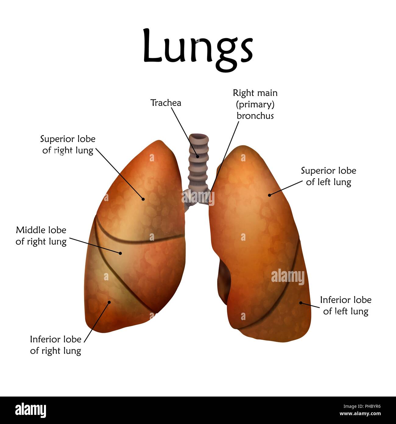
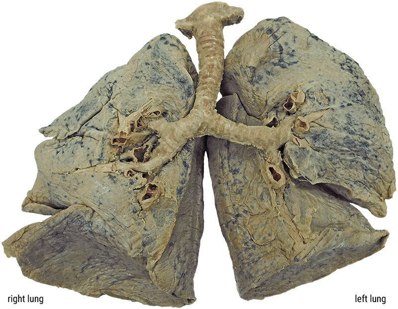
Normal chest x-ray: Anatomy tutorial | Kenhub If the lungs of a patient demonstrate diffuse markings that appear to follow the vasculature and are visible all the way to the periphery of the lungs, the patient may have vascular congestion, which can be due to heart failure. Look at the fissures as well, as these can also demonstrate thickening or excess fluid. Lung Pulmo 1/2 Lung Lobes and Fissures | GetBodySmart In the right lung, a horizontal fissure separates the superior and middle lobes and an oblique fissure separates the middle and inferior lobes. A second oblique fissure separates the two lobes of the left lung. Continue learning about the respiratory system with these quizzes and labeled diagrams. 1 2 3 4 HOW TO MAKE A LUNG MODEL WITH KIDS - hello, Wonderful Add some tape to the bottom to keep the two straws together. Step 4. Glue or double stick tape your nose and lips to the straws. Step 5. Cut the zipper part of your sandwich bags out. Step 6. Tape your lung printable to the back of the straws. Step 7. Tape your bag to each lung, tightly so no air escapes. The Lungs And Breathing Quiz! Trivia - ProProfs Quiz The lungs are one of the essential parts of your body. Lungs are necessary for life because they allow you to breathe. One of the vital roles that your lungs perform is to intake air and oxygen that your body needs to survive. Take this quiz and find out exactly just how much you know about the lungs and breathing. Questions and Answers 1.
Idiopathic Pulmonary Arterial Hypertension - What You Need to Know A V/Q scan is a two-part test which takes pictures of your lungs to look for certain lung problems. During the perfusion part of the test, radioactive dye is put into your vein (blood vessel). The blood carries the dye to the blood vessels in your lungs. Pictures are taken to see how blood flows in your lungs. Respiratory Illnesses: 13 Types of Lung Infections - OnHealth Upper Respiratory Infection. Types of upper respiratory infection include the common cold (head cold), the mild flu, tonsillitis, laryngitis, and sinus infection. Of the upper respiratory infection symptoms, the most common is a cough. Lung infections may also lead to a stuffy or runny nose, sore throat, sneezing, achy muscles, and headache. Lung - Wikipedia The lungs are located in the chest on either side of the heart in the rib cage.They are conical in shape with a narrow rounded apex at the top, and a broad concave base that rests on the convex surface of the diaphragm. The apex of the lung extends into the root of the neck, reaching shortly above the level of the sternal end of the first rib.The lungs stretch from close to the backbone in the ... NEOMED Library: Anatomical Models and Keys: Anatomy Models Heart and Lung Larynx is missing . Brain Stem Enlarged 3X. Nerves of the Head. Brain Stem (SOMSO) - Detailed. Cranial Layers . Diencephalon. Ventricles and Basal Nuclei. Muscled Arm. Muscular Leg. Skull with Facial Muscles. Foot Model . Next: Anatomy Model Keys >> Last Updated: Feb 25, 2022 4:24 PM;
Lung cancer - Symptoms and causes - Mayo Clinic Lung cancer typically doesn't cause signs and symptoms in its earliest stages. Signs and symptoms of lung cancer typically occur when the disease is advanced. Signs and symptoms of lung cancer may include: A new cough that doesn't go away. Coughing up blood, even a small amount. Shortness of breath.
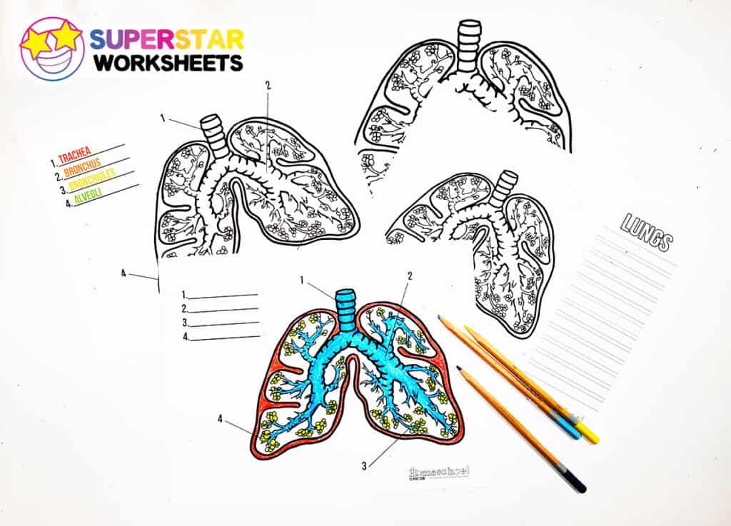
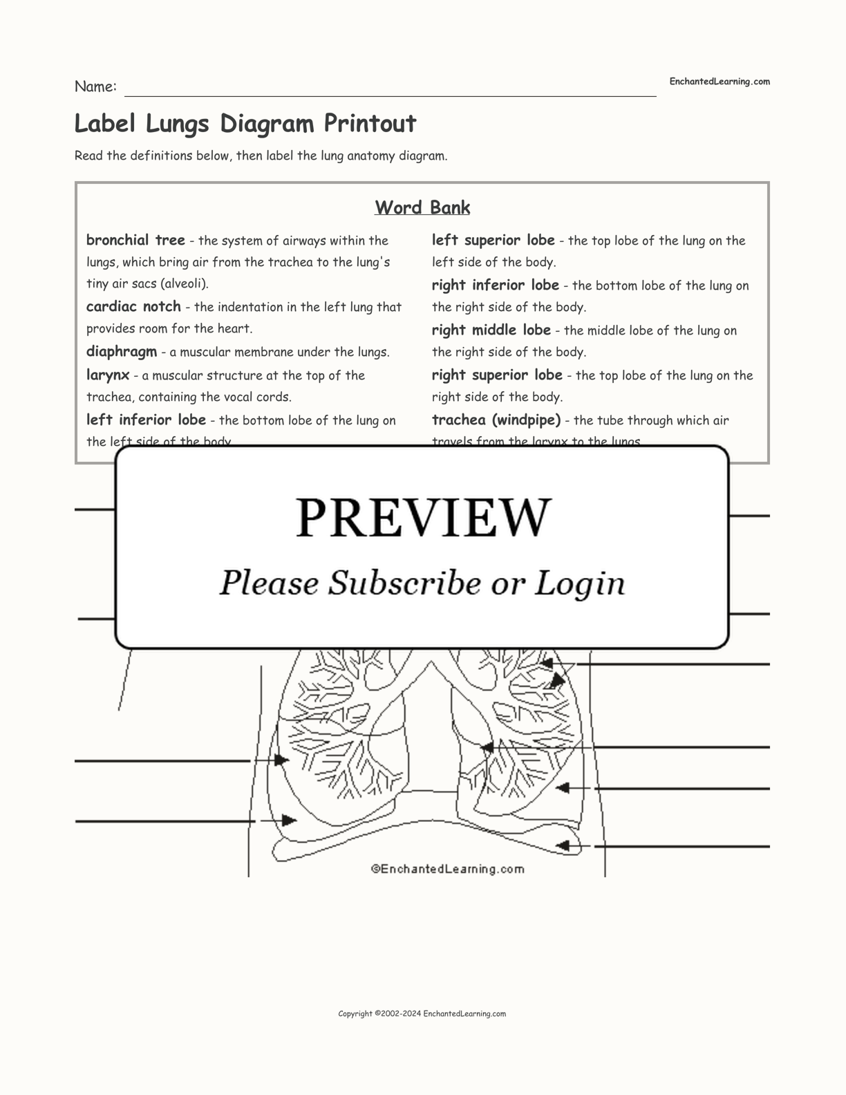
Free Respiratory System Worksheets and Printables - Homeschool Giveaways Respiratory System Doodle Labeled Coloring Page - This coloring page includes wonderful details about the respiratory system such as an explanation about how the diaphragm contracts and a close-up image of the lung alveoli. If your kids love to color, this is the perfect worksheet for you! Respiratory System Notebooking Pages
Pictures of Tips for Living With Pulmonary Arterial Hypertension - WebMD Keep your go-to kitchen items on the countertop. Put laundry soap next to or just above the machine. Move your bathroom products from under the sink up to the medicine cabinet. You want your chest ...
How the Lungs Work - The Lungs | NHLBI, NIH - National Institutes of Health The Lungs. Your lungs are the pair of spongy, pinkish-gray organs in your chest. When you inhale (breathe in), air enters your lungs, and oxygen from that air moves to your blood. At the same time, carbon dioxide, a waste gas, moves from your blood to the lungs and is exhaled (breathed out). This process, called gas exchange, is essential to life.
Body Cavities Labeled: Organs, Membranes, Definitions, Diagram ... - EZmed The right lung is highlighted in red with the right pleural cavity surrounding it shown in yellow below. The left lung is uncolored as a reference. Each pleural cavity contains a small amount of fluid, called pleural fluid. The pleural fluid helps lubricate the membranes lining the pleural cavity and lungs when breathing in and out.
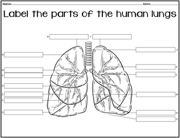
Respiratory system quizzes and labeled diagrams | Kenhub Take a look at the labeled diagram of the respiratory system above. As you can see, there are several structures to learn. Spend a few minutes reviewing the name and location of each one, then try testing your knowledge by filling in your own diagram of the respiratory system (unlabeled) using the PDF download below. Respiratory system unlabeled
human respiratory system | Description, Parts, Function, & Facts human respiratory system, the system in humans that takes up oxygen and expels carbon dioxide. The human gas-exchanging organ, the lung, is located in the thorax, where its delicate tissues are protected by the bony and muscular thoracic cage. The lung provides the tissues of the human body with a continuous flow of oxygen and clears the blood of the gaseous waste product, carbon dioxide.
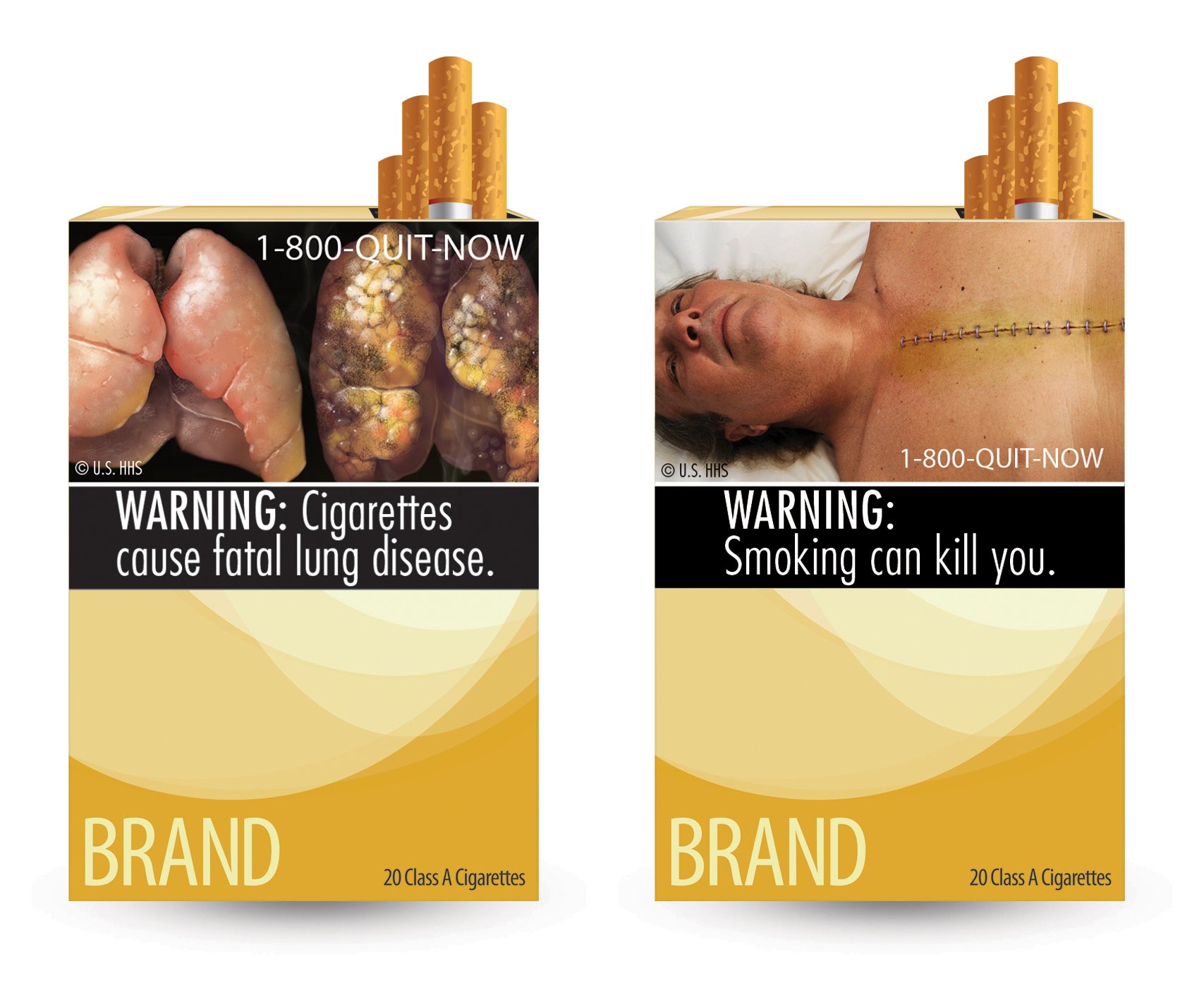
Smoker's Lungs vs. Normal Healthy Lungs - Verywell Mind Pictorial warnings, or images of lungs damaged by smoking, can be effective in preventing people from buying cigarettes. The Food and Drug Administration (FDA) has made efforts to list the health risks of tobacco use and place graphic images—such as damaged lungs—onto cigarette packaging in an effort to curb smoking. 1 How Smoking Changes the Lungs
Anatomical Planes of Body | What Are They?, Types & Position In Body lungs and organs within this ventral cavity is known as viscera. Vertebral Cavity. The posterior portion of the dorsal cavity within the vertebral column is known as the vertebral cavity. Among all body cavities, it is the narrower body cavity and seems like a thread. It is filled with the spinal cord, meninges of the spinal cord, and left ...
Phlegm colors and textures, treatment, and when to seek care Green. Green phlegm indicates a widespread and robust immune response. The white blood cells, germs, and other cells and proteins that the body produces during the immune response give the phlegm ...
Alveoli: Function, Structure, and Lung Disorders - Verywell Health Alveoli are tiny, balloon-shaped air sacs in your lungs. The function of the alveoli is to move oxygen and carbon dioxide (CO2) molecules into and out of your bloodstream. Alveoli are an important part of your respiratory system, which includes the parts of your body that help you breathe . This article discusses the structure and function of ...
Free Heart Worksheets for Human Anatomy Lessons - Homeschool Giveaways Print out sheet of the human heart with labels - This fun heart worksheet shows kids the different parts of the heart. They'll learn about the left ventricle, the left atrium, the tricuspid valve, and more. Human Heart Clipart - There is a coloring page, heart labeling worksheet and heart anatomy chart.
Smoker's Lungs Pictures | New Health Advisor 5 Smokers Lungs Pictures That Will Shock You If you have a weak stomach, you may not want to look at the following pictures. If you are a smoker, you will want to stop. Image 1: Lung of People Smoking for 15 Years The no.1 image is that of someone who has been smoking for 15 years. As you can see the lung has been covered in black spots.
Lung Volumes - Definitions - Measuring - TeachMePhysiology It is useful to divide the total space within the lungs into volumes and capacities. These can be measured to aid in the definitive diagnosis, quantification and monitoring of disease. They allow an assessment of the mechanical condition of the lungs, its musculature, airway resistance and the effectiveness of gas exchange at the alveolar membrane while being, for the most part, cheap, non ...





:background_color(FFFFFF):format(jpeg)/images/library/9673/lungs-in-situ_english.jpg)





![MCQ] - Carefully study the diagram of the human respiratory ...](https://d1avenlh0i1xmr.cloudfront.net/504ac16c-f16c-4843-bc88-0a8a95e1521f/q11---human-respiratory-system-with-labels---teachoo.jpg)


Post a Comment for "39 lungs pictures with labels"