43 cell structure with labels
Plant Cell - Definition, Structure, Function, Diagram & Types The plant cell wall is also involved in protecting the cell against mechanical stress and providing form and structure to the cell. It also filters the molecules passing in and out of it. The formation of the cell wall is guided by microtubules. It consists of three layers, namely, primary, secondary and the middle lamella. Animal Cells: Labelled Diagram, Definitions, and Structure The endoplasmic reticulum (s) are organelles that create a network of membranes that transport substances around the cell. They have phospholipid bilayers. There are two types of ER: the rough ER, and the smooth ER. The rough endoplasmic reticulum is rough because it has ribosomes (which is explained below) attached to it.
Labeled Plant Cell With Diagrams | Science Trends The parts of a plant cell include the cell wall, the cell membrane, the cytoskeleton or cytoplasm, the nucleus, the Golgi body, the mitochondria, the peroxisome's, the vacuoles, ribosomes, and the endoplasmic reticulum. Parts Of A Plant Cell The Cell Wall Let's start from the outside and work our way inwards.
Cell structure with labels
A Labeled Diagram of the Plant Cell and Functions of its Organelles A Labeled Plant Cell Amyloplasts A major component of plants that are starchy in nature, the amyloplasts are organelles that store starch. They are classified as plastids, and are also known as starch grains. They are responsible for the conversion of starch into sugar, that gives energy to the starchy plants and tubers. Cell Organelles- Definition, Structure, Functions, Diagram In a plant cell, the cell wall is made up of cellulose, hemicellulose, and proteins while in a fungal cell, it is composed of chitin. A cell wall is multilayered with a middle lamina, a primary cell wall, and a secondary cell wall. The middle lamina contains polysaccharides that provide adhesion and allow binding of the cells to one another. Structure of Cell: Definition, Types, Diagram, Functions - Embibe Cells are the fundamental structural and functional unit of all living beings including plants, animals and microorganisms. All living organisms in this universe are made up of cells. We cannot see cells with naked eyes as they are only \ (10\) microns in size whereas human eyes cannot see objects less than \ (100\) microns.
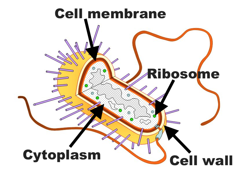
Cell structure with labels. Bacteria Cell Structures with Labels Stock Vector - Dreamstime Bacteria Cell Structures with labels. Royalty-Free Vector. Bacterial cell structures labeled on a bacillus cell with nucleoid DNA and ribosomes. External structures include the capsule, pili, and flagellum. Morphology of internal structures of bacteria. cell anatomy bacteria, en.wikipedia.org › wiki › History_of_cell_membraneHistory of cell membrane theory - Wikipedia In this view, the cell was seen to be enclosed by a thin surface, the plasma membrane, and cell water and solutes such as a potassium ion existed in a physical state like that of a dilute solution. In 1889, Hamburger used hemolysis of erythrocytes to determine the permeability of various solutes. By measuring the time required for the cells to ... Label Cell Parts | Plant & Animal Cell Activity | StoryboardThat Create a cell diagram with each part of plant and animal cells labeled. Include descriptions of what each organelle does. Click "Start Assignment". Find diagrams of a plant and an animal cell in the Science tab. Using arrows and Textables, label each part of the cell and describe its function. Plant Cells: Labelled Diagram, Definitions, and Structure The cell wall is made of cellulose and lignin, which are strong and tough compounds. Plant Cells Labelled Plastids and Chloroplasts Plants make their own food through photosynthesis. Plant cells have plastids, which animal cells don't. Plastids are organelles used to make and store needed compounds. Chloroplasts are the most important of plastids.
Structure of Bacterial Cell (With Diagram) - Biology Discussion Cell wall: It is a tough and rigid structure of peptidoglycan with accessory specific materials (e.g. LPS, teichoic acid etc.) surrounding the bacterium like a shell and lies external to the cytoplasmic membrane. It is 10-25 nm in thickness. It gives shape to the cell. Nucleus: The single circular double-stranded chromosome is the bacterial genome. 03 Label the Cell Diagram | Quizlet 03 Label the Cell STUDY Learn Flashcards Write Spell Test PLAY Match Gravity Created by muskopf1TEACHER Terms in this set (14) Nucleus Control center of the cell Nucleolus Ribosome synthesis Rough Endoplasmic Reticulum Protein transport Smooth Endoplasmic Reticulum Lipid synthesis Mitochondrion Cellular Respiratoin Golgi Apparatus Plant Cell- Definition, Structure, Parts, Functions, Labeled Diagram Plant cells are eukaryotic cells, that are found in green plants, photosynthetic eukaryotes of the kingdom Plantae which means they have a membrane-bound nucleus. They have a variety of membrane-bound cell organelles that perform various specific functions to maintain the normal functioning of the plant cell. Structure of Plant cell Cell: Structure and Functions (With Diagram) - Biology Discussion Eukaryotic Cells: 1. Eukaryotes are sophisticated cells with a well defined nucleus and cell organelles. 2. The cells are comparatively larger in size (10-100 μm). 3. Unicellular to multicellular in nature and evolved ~1 billion years ago. 4. The cell membrane is semipermeable and flexible. 5. These cells reproduce both asexually and sexually.
byjus.com › biology › diagram-of-neuronDiagram Of Neuron with Labels - BYJUS A neuron is a specialized cell, primarily involved in transmitting information through electrical and chemical signals. They are found in the brain, spinal cord and the peripheral nerves. A neuron is also known as the nerve cell. The structure of a neuron varies with their shape and size and it mainly depends upon their functions and their ... › books › NBK26880Looking at the Structure of Cells in the Microscope A typical animal cell is 10–20 μm in diameter, which is about one-fifth the size of the smallest particle visible to the naked eye. It was not until good light microscopes became available in the early part of the nineteenth century that all plant and animal tissues were discovered to be aggregates of individual cells. Chromosome Structure (Labeling) - The Biology Corner Chromosome Structure. Shannan Muskopf June 3, 2019. This simple worksheet shows a diagram of a chromosome and where it is located in the nucleus of the cell. Students use a word bank to label the chromatid, centromere, chromosomes, cell membrane, DNA, and nucleus. This worksheet was created for introductory biology for students to practice ... Labeled Cell - an overview | ScienceDirect Topics Labeled Cell. The labeled cells show a different color from the background, which makes the cell easy to be segmented. From: Control Systems Design of Bio-Robotics and Bio-mechatronics with Advanced Applications, 2020. ... So the structures of TO and its derivatives are not degraded, and the dyes could penetrate through the cell membrane into ...
Cell structures- no labels Diagram | Quizlet A. Maple trees are introduced to the land, where they grow well. B. Human activities drain about half of the water that the land generally receives. C. A beetle that infects ash trees is introduced to the land. D. About one third of the ash trees are cut down and hauled away. E. About one third of the land is cleared of all trees and used for ...
Plant and Animal Cell: Labeled Diagram, Structure, Function - Embibe Double membrane-bound structures found only in the plant cells. 2. This is an autonomous organelle. 3. There are stroma or matrix and grana or stacked discs that are involved in photosynthesis. 4. Grana are the site for photochemical reactions of photosynthesis, while stroma is the site for biochemical reactions of photosynthesis.
Cell - Label | Cell Structure Quiz - Quizizz answer choices Cell wall Cell membrane Nuclear membrane Gatekeeper Question 3 30 seconds Q. Label #5 answer choices Nucleus Nucleolus Endoplasmic Reticulum Mitochondria Question 4 30 seconds Q. Label #6 answer choices Mitochondria Chromatins Golgi Bodies Ribosomes Question 5 30 seconds Q. Label #7 answer choices Lysosomes Golgi Bodies Nucleus
Animal Cell - Structure, Function, Diagram and Types The cell is the structural and functional unit of life. These cells differ in their shapes, sizes and their structure as they have to fulfil specific functions. Plant cells and animal cells share some common features as both are eukaryotic cells. However, they differ as animals need to adapt to a more active and non-sedentary lifestyle.
Animal Cell Diagram with Label and Explanation: Cell Structure, Functions Ans. Cells make up tissues, like connective tissue, skeletal tissue, nervous tissue and fatty tissue. Tissues make up organs like your heart, your liver, your brain, spleen, stomach and so on. With no cells, there are no tissues or organs. Humans would not exist. If there are no cells, life is not possible on earth. Ques.

Animal Cells Diagram with Labels Awesome Animal Cell Diagrams Labeled | Animal cell project ...
webaim.org › techniques › tablesWebAIM: Creating Accessible Tables - Data Tables Sep 18, 2017 · In this example, the "by birth" row header has a scope of row, as do the headers with the names. The cell showing the age for Jackie will have 3 headers - one column header ("Age") and two row headers ("by birth" and "Jackie"). A screen reader would identify all of them, including the data cell content (e.g., it might read "by birth. Jackie ...
File:Plant cell structure svg labels.svg - Wikipedia File:Plant cell structure svg labels.svg. Size of this PNG preview of this SVG file: 649 × 475 pixels. Other resolutions: 320 × 234 pixels | 640 × 468 pixels | 1,024 × 749 pixels | 1,280 × 937 pixels | 2,560 × 1,874 pixels. Original file (SVG file, nominally 649 × 475 pixels, file size: 113 KB) This is a file from the Wikimedia Commons.
Cell Structure and Function Quiz - Mr. Skerrett Cell Structure and Function Quiz. Cell Structure and Function Quiz Identify the following : Label 1 is. -- Choose an answer -- Cell Membrane Unit Membrane Cell Wall Cytoplasm. Label 2 is. -- Choose an answer -- Lysosome Golgi Apparatus Smooth ER Rough ER. Label 3 is.
Bacteria in Microbiology - shapes, structure and diagram The bacteria shapes, structure, and labeled diagrams are discussed below. Sizes The sizes of bacteria cells that can infect human beings range from 0.1 to 10 micrometers. Some larger types of bacteria such as the rickettsias, mycoplasmas, and chlamydias have similar sizes as the largest types of viruses, the poxviruses.
physics.bu.edu › ~okctsui › PY543Crystal Structure Basic Concepts - Boston University Unit cell and lattice constants: A unit cell is a volume, when translated through some subset of the vectors of a Bravais lattice, can fill up the whole space without voids or overlapping with itself. The conventional unit cell chosen is usually bigger than the primitive cell in favor of preserving the symmetry of the Bravais lattice. For ...
Cell Structure With Labels Teaching Resources | Teachers Pay Teachers Students BUILD either the plant or animal cell with organelles and label it. Labels include name and function of the organelle. Also included is a question sheet that can be used as a mini-assessment. Please check out other products in my store.
PDF Human Cell Diagram, Parts, Pictures, Structure and Functions The endoplasmic reticulum(ER) is a membranous structure that contains a network of tubules and vesicles. Its structure is such that substances can move through it and be kept in isolation from the rest of the cell until the manufacturing processes conducted within are completed.
Plant Cell Structure and Parts Explained With a Labeled Diagram Different Parts of a Plant Cell. Plant cells are classified into three types, based on the structure and function, viz. parenchyma, collenchyma and sclerenchyma. The parenchyma cells are living, thin-walled and undergo repeated cell division for growth of the plant. They are mostly present in the leaf epidermis, stem pith, root and fruit pulp.
› cell › fulltextIdentifying Medical Diagnoses and Treatable Diseases ... - Cell Feb 22, 2018 · The implementation of clinical-decision support algorithms for medical imaging faces challenges with reliability and interpretability. Here, we establish a diagnostic tool based on a deep-learning framework for the screening of patients with common treatable blinding retinal diseases.
Healthhype.com | Current Health Articles on Symptoms, Diseases and ... Healthhype.com | Current Health Articles on Symptoms, Diseases and ...
bioinformatics.uconn.edu › single-cell-rnaSingle-cell RNA sequencing (Cell Ranger) | Computational ... The Single Cell 3’ Protocol produces Illumina-ready sequencing libraries. A Single Cell 3’ Library comprises standard Illumina paired-end constructs which begin and end with P5 and P7. The Single Cell 3’ 16 bp 10xTM Barcode and 10 bp randomer is encoded in Read 1, while Read 2 is used to sequence the cDNA fragment.
Study the diagram. Label the structures A , B and C ... - Byju's Study the diagram. Label the structures A , B and C. What is the function of C ? Is this a plant cell or an animal cell? Draw and label 3 extra structures ...1 answer · Top answer: A Nucleus B Chloroplast C Cell wall It is a plant cell as it has a cell wall. Cell wall is a structure found only in plant cells. It supports and protect ...
Label the cell structure. | Study.com Label the cell structure. Cells: All living cells contain an intracellular space called the cytoplasm. The cytoplasm is filled with a jelly-like fluid where many of the cells enzymatic reactions...


Post a Comment for "43 cell structure with labels"